Colonoscopy: preparation, pain, sedation — what actually happens
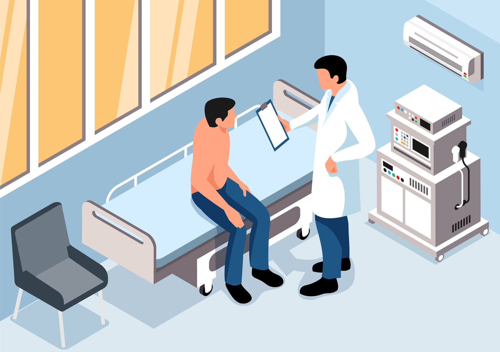
Most people postpone colonoscopy for two reasons: they fear it will hurt, and they fear the preparation. Of the two, only the second fear is justified.
In a study of 35,216 patients published in 2018, 14.2% of those examined without sedation felt pain. That is, nearly seven out of eight people had no pain even with no sedation at all. With sedation the figures fall substantially: patients under conscious sedation reported pain in 50% of cases, against only 20% under deep sedation, and moderate discomfort was 33% against 0%.
The unpleasant part, the one almost every patient talks about afterwards, is the preparation the day before.
Why it hurts, when it hurts
Pain in colonoscopy does not come from touching the mucosa — the bowel has no pain receptors of that kind. It comes from two mechanical sources:
Loops. The colon is not a straight tube. The advancing colonoscope forms loops that stretch the mesentery, and that stretching is felt. An experienced endoscopist spends a good part of the procedure undoing loops, precisely to avoid this.
Insufflation. To see the wall, the bowel has to be distended with gas. Insufflated air is absorbed slowly and produces bloating and cramps afterwards. Carbon dioxide is absorbed roughly 150 times faster and significantly reduces post-procedure discomfort. It is worth asking whether the unit uses CO2.
Sedation: the options
No sedation. Possible, and tolerated by many. Advantage: you leave immediately and can drive.
Conscious sedation. A sedative and an analgesic; you stay awake but relaxed, and usually remember little.
Deep sedation (propofol). You sleep through the procedure. The data show the best comfort of the three. It requires anaesthetic monitoring and you cannot drive afterwards.
There is no universally correct choice. If you have had a difficult colonoscopy before, if you have adhesions after abdominal surgery, or if you are very anxious, deep sedation is reasonable.
Preparation: this is where the examination is won or lost
This part is not an administrative formality. Poor preparation means a poor examination, and a poor examination means missed lesions and a repeat procedure.
A few things that matter more than they appear:
Split dosing. The cleansing solution given in two parts — some the evening before, some on the morning of the examination — cleans significantly better than the whole quantity the night before. If you are told otherwise, ask why.
Diet in the preceding days. Two to three days beforehand, drop seeds, pips, skins, wholegrains and coarse-fibre vegetables. They stay in the colon and coat the mucosa.
Fluids. The preparation dehydrates you. The permitted clear fluids have to actually be drunk, not merely listed on the sheet.
Anticoagulants and antiplatelet drugs. Discuss these in advance with the doctor who prescribed them; do not stop them on your own initiative.
Oral iron is stopped about a week beforehand, because it blackens and sticks to the mucosa.
What is seen in the rectum — and why it matters especially
This is where colonoscopy becomes relevant to rectal cancer in particular.
The rectum is the last portion examined on withdrawal and, paradoxically, can be the most deceptive. The last few centimetres, just above the anal canal, are poorly seen looking straight ahead. That is why the endoscopist performs rectal retroflexion — turning the tip of the instrument through 180 degrees to look back towards the anal canal. Lesions sited exactly in that zone can be missed without this manoeuvre.
The second rectum-specific point: distance. When a rectal tumour is found, the distance from its lower edge to the anal verge is one of the most important numbers in the whole file, because it directly influences whether the sphincter can be preserved. That distance is measured more reliably by rigid rectoscopy than by a flexible colonoscope, which can loop and falsify the measurement.
Why MRI follows colonoscopy
Colonoscopy sees the surface. It shows that a tumour exists, where it is and what it looks like, and allows the biopsy that establishes the diagnosis. What it cannot do is say how deeply the tumour has penetrated the wall, whether lymph nodes are involved, or how close the tumour lies to the mesorectal fascia.
All of that is seen on pelvic MRI, which is the examination that actually decides treatment. In rectal cancer, an operation planned without MRI is an operation planned blind.
That is why the correct sequence is: colonoscopy with biopsy, then pelvic MRI, then thoraco-abdominal CT, then the decision in a multidisciplinary meeting.
Risks
Colonoscopy is a safe procedure, but not free of risk. The relevant ones are bleeding — particularly after removal of a large polyp — and perforation, which is rare but real. Risk rises with age, comorbidity, post-surgical adhesions and the complexity of the polyps removed.
These risks have to be weighed against what the procedure offers: the only examination that prevents cancer rather than merely detecting it, because the polyp is removed in the same session in which it is found.
Afterwards
You will be bloated for a few hours — less so if CO2 was used. If you had deep sedation, do not drive that day and do not sign important documents.
Always ask for two things before you leave: the written report, stating whether the examination was complete to the caecum and how the preparation was, and the interval to your next colonoscopy. The second is the most commonly forgotten and the one that matters long term.
If biopsies were taken or polyps removed, the histology result comes in one to two weeks. That, and not the impression during the examination, is what establishes the diagnosis.
The information in this article is educational and does not replace the instructions of the doctor performing the procedure.