Pelvic MRI in rectal cancer: why it is essential for diagnosis
Pelvic magnetic resonance imaging (MRI) is considered the essential investigation in the evaluation of rectal cancer. Without a good-quality pelvic MRI, the medical team cannot plan treatment correctly. This article explains why pelvic MRI is so important and what you should know before having this scan.
Why pelvic MRI is indispensable
Pelvic MRI provides information that no other investigation can give with the same precision:
1. Assessing the depth of tumor invasion (T)
MRI shows how deep the tumor has invaded the rectal wall — whether it is limited to the mucosa, has reached the muscle layer, or has crossed the rectal wall and invades the surrounding fatty tissue (mesorectum).
2. Lymph node assessment (N)
It identifies suspicious lymph nodes in the mesorectum and beyond. Suspicion criteria include size, shape, and signal heterogeneity.
3. Relationship to the mesorectal fascia (MRF)
One of the most important pieces of information that MRI provides is the distance between the tumor and the mesorectal fascia — the envelope surrounding the mesorectum. If the tumor is less than 1 mm from this fascia, the surgical resection margin is threatened and neoadjuvant treatment becomes necessary.
4. Extramural vascular invasion (EMVI)
MRI can detect whether the tumor has invaded blood vessels outside the rectal wall, an important negative prognostic factor.
5. Assessing response to treatment
After chemoradiotherapy, a second MRI (restaging) shows how much the tumor has shrunk and helps in surgical decision-making.
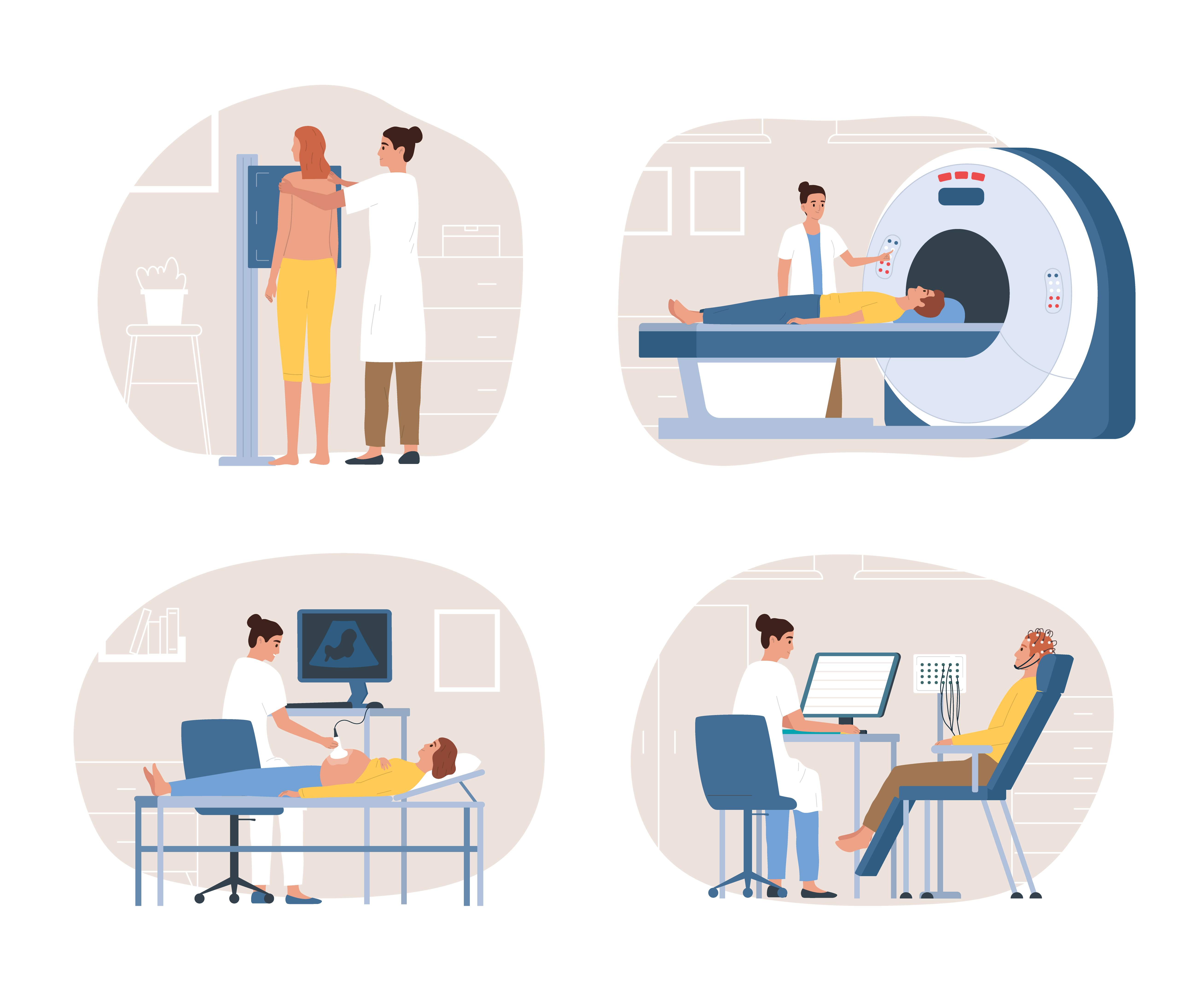
How a pelvic MRI is performed
Preparation
- No special preparation required — you do not need to fast
- Inform the doctor if you have metal implants, a pacemaker or claustrophobia
- An antispasmodic medication (Buscopan) may be given to reduce bowel movements during the scan
Procedure
- Duration: 30-45 minutes
- You will lie on your back inside the scanner tunnel
- It is a painless investigation with no ionizing radiation
- The machine produces loud noises — you will be given headphones or earplugs
- It is important to stay as still as possible for high-quality images
After the scan
- You can immediately return to normal activities
- Results are interpreted by a specialist radiologist
- The detailed report is sent to your doctor in a few days
What the MRI report contains
A high-quality MRI report for rectal cancer should include:
- Tumor location — distance from the anal verge, circumferential position
- T stage — depth of invasion
- N stage — number and characteristics of suspicious nodes
- Distance to the mesorectal fascia — in millimeters
- Presence of EMVI — extramural vascular invasion
- Relationship to the anal sphincter — important for the decision on sphincter preservation
- mrTNM stage — complete staging based on MRI
Difference between MRI and other investigations
| Investigation | What it shows best | Limitations |
|---|---|---|
| Pelvic MRI | Local extent, mesorectal fascia, EMVI | Does not detect distant metastases |
| Chest-abdomen CT | Metastases (liver, lungs) | Limited precision for local T stage |
| Endorectal ultrasound | Superficial tumors (T1-T2) | Limited for advanced tumors |
| PET-CT | Metabolic activity, occult metastases | Does not replace MRI for local staging |
Frequently asked questions
Do I need an MRI before surgery?
Yes, always. ESMO and NCCN guidelines mandate pelvic MRI before any therapeutic decision in rectal cancer. Without MRI, the surgeon cannot plan the operation correctly.
Can I have an MRI if I am claustrophobic?
Yes. Your doctor can prescribe a mild sedative before the procedure. Some centers also have open-bore MRI machines.
How accurate is MRI?
Pelvic MRI has an accuracy of 85-90% for T stage and 75-80% for N stage. It is the best investigation available for local evaluation of rectal cancer.
Do I need to repeat the MRI after chemoradiotherapy?
Yes. A restaging MRI is essential 6-8 weeks after the end of radiotherapy to assess tumor response and guide the surgical decision.
This article is for informational purposes only and does not replace medical consultation. Discuss with your doctor about which investigations you need.